MRI

MRI

The Elliot has state-of-the-art, open MRI machines at three convenient locations. Our imaging provides an excellent way to diagnose disease, injuries, infections, and inflammatory conditions affecting the body. We have technology for metal suppression, advanced abdominal capabilities and a variety of ways to ease claustrophobia.

About

Magnetic Resonance Imaging (MRI) is an advanced medical imaging technique that produces cross-sectional images of the body without using radiation. The images are produced using a large superconducting magnet, radio frequency waves, and a computer system.  The MRI machine uses the water molecules in your body and an applied Radiofrequency wave. Your molecules act like tiny magnets and our cameras act like antennas to receive signal. This signal is collected and are processed by powerful computers to generate images. When the Radiofrequency is applied, gradient pulses cause a very loud sound.

Click here to hear a sample of the noises our machine makes.

Risks

A thorough screening process is completed on all patients to determine if it is safe for you to have an MRI. While there are many metal implants that are perfectly safe in the MRI environment, there are some implants that should not be scanned or may require certain precautions.

It is very important to communicate all your implant information to the MRI team. Please bring any implant cards with you to your appointment.

Only MRI Conditional pacemakers or Defibrillators can be scanned under certain circumstances. A coordinated MRI appointment can be made after research and Cardiology orders have been completed.

As implants are affected by the magnetic field used in an MRI exam, special precautions must be followed. Please let the MRI team know if you have any of the following:

  • Cardiac Pacemaker/ defibrillator
  • Aneurysm clips
  • Spinal Stimulator
  • Diabetic Pump
  • Breast Tissue Expander
  • Harrington Rods
  • Metal plates, pins, or other metallic implants
  • Shrapnel from a gunshot wound
  • Ever been a metal worker
  • Permanent tattoo eyeliner
  • Cochlea implant

Preparation

Before an MRI exam, you may eat and take your medications as normal unless otherwise instructed. Depending on which part of the body is being examined, you may be asked to remove some or all of your clothing, as well as any items that might affect the magnetic imaging, like jewelry, eyeglasses, hearing aids, underwire bras, etc. During your MRI, you will be given hearing protection, as the MRI machine makes a series of loud noises. Click here to listen

Many patients struggle with claustrophobia. Our staff are experienced and have several methods to help you through the exam. Some patients opt to take medication for claustrophobia or anxiety during the exam. If you intend to take medication, please get in touch with your provider before your MRI. Do not take your medication until you arrive for the MRI, and the technologist has instructed you to do so.

Click on any exam below to explore in better detail what to expect and how to prepare for your MRI at The Elliot.

What to Expect

If you have been booked for an MRI of your spine (cervical, thoracic, lumbar) you will be asked to arrive at the MRI department 15 minutes prior to your appointment time. If the Doctor ordering your exam has requested contrast, we ask you to arrive 30 minutes prior to your appointment time.

After the safety screening has been completed you will be provided a locker where you can safely lock your valuables. We recommend leaving as many personal belongings at home as possible. MRI Safe clothing will be provided for you.

In the scan room you will be asked to lie on your back onto an MRI coil (camera). You will be given hearing protection as the MRI machine makes a series of loud noises.

You will also be given the option of listening to music if you would like.

The length of a spine MRI is typically 15-25 minutes without a contrast injection. The total length of time is 30-35 minutes if a contrast injection is needed.

You will be asked to hold still as MRI scans are sensitive to motion and any motion may obscure information the Radiologist uses to make a formal report.

Your referring physician will explain this formal report to you.

If you have been booked for an MRI of your brain you will be asked to arrive at the MRI department 15 minutes prior to your appointment time. If the Doctor ordering your exam has requested contrast, we ask you to arrive 30 minutes prior to your appointment time.

After the safety screening has been completed you will be provided a locker where you can safely lock your valuables. We recommend leaving as many personal belongings at home as possible. MRI Safe clothing will be provided for you.

In the scan room you will be asked to lie on your back onto an MRI coil (camera) and an additional coil will be clicked in place acting as an antenna. This device has prism technology allowing you to see outside of the machine, either looking at the technologists or the picture window depending on the site you are being scanned at. You will be given hearing protection as the MRI machine makes a series of loud noises.

You will also be given the option of listening to music if you would like.

The length of a brain MRI is typically 20-25 minutes without a contrast injection. The total length of time is 30-35 minutes if a contrast injection is needed. You will be asked to hold still as MRI scans are sensitive to motion and any motion may obscure information the Radiologist uses to make a formal report.

Your referring physician will explain this formal report to you.

If you have been booked for an orbit MRI you will be asked to arrive at the MRI department 15 minutes prior to your appointment time. If the Doctor ordering your exam has requested contrast, we ask you to arrive 30 minutes prior to your appointment time.

After the safety screening has been completed you will be provided a locker where you can safely lock your valuables. We encourage you to leave as many valuables as possible at home. MRI Safe clothing will be provided for you.

In the scan room you will be asked to lie on your back onto an MRI coil (camera) and an additional coil will be clicked in place acting as an antenna. You will be provided with an eye mask to prevent motion of the eyes during this scan.

You will be given hearing protection as the MRI machine makes a series of loud noises.

You will also be given the option of listening to music if you would like.

The length of a brain MRI is typically 20-25 minutes without a contrast injection. The total length of time is 30-35 minutes if a contrast injection is needed.

A formal report of the results will be sent to your referring physician who will explain the results to you.

If you have been booked for an MRI of your chest you will be asked to arrive at the MRI department 15 minutes prior to your appointment time. If the Doctor ordering your exam has requested contrast, we ask you to arrive 30 minutes prior to your appointment time.

After the safety screening has been completed you will be provided a locker where you can safely lock your valuables however we encourage you to leave as many valuables as possible at home. MRI Safe clothing will be provided for you.

Your test may require an injection of MRI contrast. The Elliot MRI department uses Gadavist and requires an IV be started in either your hand or your arm.

In the scan room you will be asked to lie on your back onto an MRI coil (camera). An additional camera is placed over the chest acting as an antenna.

You will be given hearing protection as the MRI machine makes a series of loud noises.

You will also be given the option of listening to music if you would like.

The length of a chest MRI is typically 30-35 minutes and will require some scans where we will instruct you to hold your breath.

If you have been booked for an MRI of your pelvis you will be asked to arrive at the MRI department 15 minutes prior to your appointment time. If the Doctor ordering your exam has requested contrast, we ask you to arrive 30 minutes prior to your appointment time.

After the safety screening has been completed you will be provided a locker where you can safely lock your valuables. We encourage you to leave as many valuables as possible at home. MRI Safe clothing will be provided for you.

In the scan room you will be asked to lie on your back onto an MRI coil (camera) and an additional coil will be placed over your pelvis and acts as an antenna. You will have the option to go into the MRI feet first if you would feel more comfortable this way.

You will be given hearing protection as the MRI machine makes a series of loud noises.

You will also be given the option of listening to music if you would like.

The length of a pelvis MRI can vary depending on the indication. Pelvis MRI without contrast is typically 20-25. When contrast is used the total scan time averages between 35-45 minutes. A formal report of the results will be sent to your referring physician who will explain the results to you.

If you have been booked for an MRI of your hip you will be asked to arrive at the MRI department 15 minutes prior to your appointment time. If the Doctor ordering your exam has requested contrast, we ask you to arrive 30 minutes prior to your appointment time.

After the safety screening has been completed you will be provided a locker where you can safely lock your valuables. We encourage you to leave as many valuables as possible at home. MRI Safe clothing will be provided for you.

In the scan room you will be asked to lie on your back onto an MRI coil (camera) and an additional coil will be placed over your pelvis and acts as an antenna. You will have the option to go into the MRI feet first if you would feel more comfortable this way.

You will be given hearing protection as the MRI machine makes a series of loud noises.

You will also be given the option of listening to music if you would like.

The length of a hip MRI can vary depending on the indication. Hip MRI without contrast is typically 20-25 minutes and 30-45 minutes when contrast is needed.

A formal report of the results will be sent to your referring physician who will explain the results to you.

If you have been booked for an MRI of your knee you will be asked to arrive at the MRI department 15 minutes prior to your appointment time. If the Doctor ordering your exam has requested contrast, we ask you to arrive 30 minutes prior to your appointment time.

After the safety screening has been completed you will be provided a locker where you can safely lock your valuables. We encourage you to leave as many valuables as possible at home. MRI Safe clothing will be provided for you.

In the scan room you will be asked to lie on your back onto an MRI coil (camera). An additional camera is placed over the affected knee acting as an antenna. This exam is done feet first and your head will not go into the scanner.

You will be given hearing protection as the MRI machine makes a series of loud noises.

You will also be given the option of listening to music if you would like.

The length of a knee MRI is typically 20-25 minutes and will require you to hold very still. MRI scans are sensitive to motion and any motion may obscure information the Radiologist uses to make a formal report.

The formal report of the results will be sent to your referring physician who will explain the results to you.

If you have been booked for an MRI of your ankle you will be asked to arrive at the MRI department 15 minutes prior to your appointment time. If the Doctor ordering your exam has requested contrast, we ask you to arrive 30 minutes prior to your appointment time.

After the safety screening has been completed you will be provided a locker where you can safely lock your valuables. We encourage you to leave as many valuables as possible at home. MRI Safe clothing will be provided for you.

In the scan room you will be asked to lie on your back onto an MRI coil (camera). An additional camera is placed over the affected ankle acting as an antenna. This exam is done feet first and your head will not go into the scanner.

You will be given hearing protection as the MRI machine makes a series of loud noises.

You will also be given the option of listening to music if you would like.

The length of an ankle MRI is typically 30-35 minutes and will require you to hold very still. MRI scans are sensitive to motion and any motion may obscure information the Radiologist uses to make a formal report.

If you have been booked for an MRI of your foot you will be asked to arrive at the MRI department 15 minutes prior to your appointment time. If the Doctor ordering your exam has requested contrast, we ask you to arrive 30 minutes prior to your appointment time.

After the safety screening has been completed you will be provided a locker where you can safely lock your valuables. We encourage you to leave as many valuables as possible at home. MRI Safe clothing will be provided for you.

In the scan room you will be asked to lie on your back onto an MRI coil (camera). An additional camera is placed over the affected foot acting as an antenna. This exam is done feet first and your head will not go into the scanner.

You will be given hearing protection as the MRI machine makes a series of loud noises.

You will also be given the option of listening to music if you would like.

The length of a foot MRI is typically 30-35 minutes and will require you to hold very still. MRI scans are sensitive to motion and any motion may obscure information the Radiologist uses to make a formal report.

The formal report of the results will be sent to your referring physician who will explain the results to you.

If you have been booked for an MRI of your shoulder you will be asked to arrive at the MRI department 15 minutes prior to your appointment time. If the Doctor ordering your exam has requested contrast, we ask you to arrive 30 minutes prior to your appointment time.

After the safety screening has been completed you will be provided a locker where you can safely lock your valuables. We encourage you to leave as many valuables as possible at home. MRI Safe clothing will be provided for you.

In the scan room you will be asked to lie on your back onto an MRI coil (camera). An additional camera is placed over the affected shoulder acting as an antenna. We will position your arm down by your side and will support it using pillows.

You will be given hearing protection as the MRI machine makes a series of loud noises.

You will also be given the option of listening to music if you would like.

The length of a shoulder MRI is typically 20-25 minutes and will require you to hold very still. MRI scans are sensitive to motion and any motion may obscure information the Radiologist uses to make a formal report.

The formal report of the results will be sent to your referring physician who will explain the results to you.

If you have been booked for an MRI of your elbow you will be asked to arrive at the MRI department 15 minutes prior to your appointment time. If the Doctor ordering your exam has requested contrast, we ask you to arrive 30 minutes prior to your appointment time.

After the safety screening has been completed you will be provided a locker where you can safely lock your valuables. We encourage you to leave as many valuables as possible at home. MRI Safe clothing will be provided for you.

In the scan room you will be asked to lie on your back onto an MRI coil (camera). An additional camera is placed around the elbow and acts as an antenna. This exam can be done head-first or feet first depending on your preference.

You will be given hearing protection as the MRI machine makes a series of loud noises.

You will also be given the option of listening to music if you would like.

The length of an elbow MRI is typically 20-25 minutes and will require you to hold very still. MRI scans are sensitive to motion and any motion may obscure information the Radiologist uses to make a formal report.

The formal report of the results will be sent to your referring physician who will explain the results to you.

If you have been booked for an MRI of your wrist you will be asked to arrive at the MRI department 15 minutes prior to your appointment time. If the Doctor ordering your exam has requested contrast, we ask you to arrive 30 minutes prior to your appointment time.

After the safety screening has been completed you will be provided a locker where you can safely lock your valuables. We encourage you to leave as many valuables as possible at home. MRI Safe clothing will be provided for you.

In the scan room you will be asked to lie on your back onto an MRI coil (camera). An additional camera is placed around the wrist and acts as an antenna. This exam can be done head first or feet first depending on your preference.

You will be given hearing protection as the MRI machine makes a series of loud noises.

You will also be given the option of listening to music if you would like.

The length of a wrist MRI is typically 20-25 minutes and will require you to hold very still. MRI scans are sensitive to motion and any motion may obscure information the Radiologist uses to make a formal report.

The formal report of the results will be sent to your referring physician who will explain the results to you.

If you have been booked for an MRI of your hand you will be asked to arrive at the MRI department 15 minutes prior to your appointment time.

After the safety screening has been completed you will be provided a locker where you can safely lock your valuables. We encourage you to leave as many valuables as possible at home. MRI Safe clothing will be provided for you.

In the scan room you will be asked to lie on your back onto an MRI coil (camera). An additional camera is placed around the hand and acts as an antenna. This exam can be done head first or feet first depending on your preference.

You will be given hearing protection as the MRI machine makes a series of loud noises.

You will also be given the option of listening to music if you would like.

The length of a hand MRI is typically 20-25 minutes if it is without contrast. The scan will take approximately 30-35 minutes if an injection is needed. The contrast Elliot uses for MRI is called Gadavist and requires that an IV be started in either your arm or hand. Your referring physician will determine if the injection is necessary. It is important to hold very still. MRI scans are sensitive to motion and any motion may obscure information the Radiologist uses to make a formal report.

The formal report of the results will be sent to your referring physician who will explain the results to you.

If you have been booked for an MRI of your abdomen (liver, spleen, kidneys, adrenals & pancreas) you will be asked to arrive at the MRI department 15 minutes prior to your appointment time. If the Doctor ordering your exam has requested contrast, we ask you to arrive 30 minutes prior to your appointment time.

After the safety screening has been completed you will be provided a locker where you can safely lock your valuables. We encourage you to leave as many valuables as possible at home. MRI Safe clothing will be provided for you.

Abdominal MRI requires the injection of a contrast agent. The contrast used by the Elliot is called Gadavist and requires that an IV be started in either your arm or hand.

In the scan room you will be asked to lie on your back onto an MRI coil (camera). An additional camera is placed over the abdomen and acts as an antenna.

You will be given hearing protection as the MRI machine makes a series of loud noises.

You will also be given the option of listening to music if you would like.

We will be giving you breathing instructions for this exam and will hear the technologist instruct you through the headphones.

The length of an abdomen MRI is typically 30-35 minutes and will require you to hold very still. MRI scans are sensitive to motion and any motion may obscure information the Radiologist uses to make a formal report. The formal report of the results will be sent to your referring physician who will explain the results to you.

This exam will take place at our River's Edge location on our Siemens 3T scanner.

Highly detailed images will be obtained of the prostate without the use of any invasive techniques. These images will provide mappings for future biopsy intervention if necessary.

You will be asked not to eat or drink anything for 6 hours prior to the testing as well as doing an at home bedside enema 2-3hrs prior to your appointment. This process aids in the image quality as fecal matter and gas obscure details that prevent details necessary for diagnosis.

We ask that you not wear anything containing metal, including zippers and/or articles of clothing with silver or copper threads such as antimicrobial underwear.

This exam takes 45 minutes and requires you to hold very still. You will receive an injection of contrast media that will be administered through an IV.

You will be provided with ear plugs to support hearing protection, as the machine makes a very loud knocking noise. You will also be given headphones with music for entertainment.

Please follow up with your provider if you have any questions. If you present for the MRI without this prep, the Technologist will consult with the Radiologist and the exam may be rescheduled if anatomy is obscured. 

MRI of the pelvic floor is often the only way to show how the muscles are working. This cannot be seen or determined in your doctor’s office visit. It can often be the test that provides the answer to the issues experienced over a period of time.

If you have been scheduled for a MR Defecography you will be asked to arrive 15 minutes prior to the start of your exam. After the necessary safety screening form is complete you will be asked to change into MRI Safe clothing. You will be given disposable underwear to wear as well.

You will be asked to lie on the MRI table where you will be covered up with a blanket or sheet to give you privacy.

Your privacy is always a priority.

Gel will be inserted into your rectum using a small enema tip. The gel highlights the anatomy and differentiates the muscles.

This exam will take approximately 15-20 minutes

The first pictures are obtained with you relaxing normally followed by a series of instructions including the following;

  1. Squeezing and releasing your pelvic muscles for 30 second (similar to Kegal exercises)
  2. Bearing down for 30 seconds and relaxing for 30 seconds
  3. Lastly you will be asked to attempt to push the gel out and it may or may not come out but either way it will show what your muscles are doing. Remember, you are wearing a garment and there is no worry of any gel ending up on the exam table.

The exam is now complete and you will be given the necessary items to clean up as well as additional disposable underwear to wear home.

If you have been scheduled for an MR Enterography you will be asked to arrive 1 Hour prior to the start of your exam. It is necessary to abstain from eating or drinking any food or liquid for 6 Hours prior to the time of your exam. [If you are scheduled at 8:00am you can have nothing to eat or drink after 1am and arrive at the MRI department for 7am]NO FOOD or DRINK for 6 Hours.

After the necessary safety screening form is complete you will change into MRI safe clothing.

You will be asked to drink 3 bottles of oral contrast (Breeza) which distends collapsed loops of bowel by replacing ingested food and air with a mineral to create a uniform consistency. You will need to drink 1 bottle approximately every 20 minutes.

You will be asked to lie on your belly onto a coil which is an MRI camera. An additional coil will be placed on your back. We ask you to lie on your belly in order to spread out the bowel as best possible for visualization.

This exam requires the use of a contrast agent called Gadavist. The Gadavist will be administered by using an IV in either your arm or hand.

An additional injection called Glucagon will be given by a Radiology Nurse midway through the exam. This will then be repeated toward the end of the exam. The purpose of this injection is to relax the bowel.

You may experience cramping and bloating during this procedure and are advice not to eat heavy solid food for the remainder of the day as this can cause constipation.

The scan time for this exam lasts approximately 25 minutes.

The images obtain during the MRI will be sent to a system where a Radiologist dedicated to MRI will make a dictation. These results will be explained to you by your referring physician.

Liver fibrosis can be the result of chronic liver disease and if left untreated can become cirrhosis. Cirrhosis carries many complications such as liver failure, portal hypertension, varices, hepatocellular carcinoma (HCC), and hepatic encephalopathy. Studies have proven that liver fibrosis may be reversible in the early stages and is why early detection is important. Currently, percutaneous biopsy is the standard practice for detected liver fibrosis.

MRE is an MRI-based method to test the stiffness of the liver and provides quantitative maps of tissue stiffness over large regions of the liver. This test takes only 5-10 minutes. If done in combination with a diagnostic abdomen MRI the total time is approximately 35-45 minutes.

If you have been booked for an MRI with Liver Elastography you will be asked to arrive at the MRI department 30 minutes prior to your appointment time. After the safety screening has been completed you will be provided a locker where you can safely lock your valuables. We encourage you to leave as many valuables as possible at home. MRI Safe clothing will be provided for you.

If your exam has been ordered to be done in combination with an MRI of the abdomen, we may be using an injection of contrast called Gadavist and requires that an IV be started in either your arm or hand.

In the scan room you will be asked to lie on your back onto an MRI coil (camera). An additional camera is placed over the abdomen and acts as an antenna. For the elastography specific pictures a mechanical driver (paddle like device) will be placed over the right side of your abdomen and held in place by a compression belt. This device is meant to send sound waves to your liver and determine the amount of stiffness. You will experience a vibration sensation during the scan and requires that you hold your breath.

You will be given hearing protection as the MRI machine makes a series of loud noises.

You will also be given the option of listening to satellite radio if you would like.

MRI scans are sensitive to motion and any motion may obscure information the Radiologist uses to make a formal report.

The formal report of the results will be sent to your referring physician who will explain the results to you.

If your child has been scheduled for a sedated MRI procedure, please note that several different departments are involved. Your child’s referring provider will help you set up the appointment through our central booking department. The departments involved are the MRI department, Radiology Nurses and the Anesthesia team. Once your child has been booked for their appointment you will receive several calls to coordinate your child’s care. One call will be from the MRI staff to screen your child to ensure they are safe for the MRI exam and another from pre-registration. You will also receive a call from the pre-op department to go over instructions such as nothing to eat or drink after midnight the night before the procedure. Your child will be accompanied by the anesthesia team and the Radiology nursing staff down to the MRI department. You will be welcomed to wait in the waiting room near the recovery room where your child will go directly after the MRI procedure.

Your child’s safety is the number one priority; therefore your child will be monitored during the entire time they are sedated. The anesthesia team will stay with your child at all times, even in the MRI scan room during the procedure.

Your child will need to be free from metal so please make sure they are wearing cotton clothing without sparkles, buttons, silver threading or antimicrobial material.

Please feel free to speak with the pre-op department with any specific questions you may have about the procedure or the MRI department about questions relating to imaging.

Learn More

If you have been booked for an MR ARTHROGRAM it is important to know two departments are involved.

Central booking will schedule the MRI with your Doctor’s office to coordinate the following departments;

MRI & XRAY
Once scheduled, you will be receiving a call from the MRI department to ensure you are safe to enter the magnetic environment. You also will be asked if you are on any of the following medications prior to this procedure;

  • AGGRENOX
  • ARIXTRA
  • ASPRIN
  • COUMADIN
  • ELIQUIS
  • HEPARIN
  • LOVENOX
  • PLAVIX PLETAL
  • PRADAXA
  • XARELTO

Your MRI will be scheduled ½ hour after the XRAY appointment. [If your MRI is scheduled for 9am your Arthrogram injection appointment will be for 8:30am].

During your appointment in the XRAY department the Radiologist or Physician Assistant will be administering a small amount of contrast (Gadavist) into the joint space being imaged with the guidance of XRAY.

Following the injection is your MRI appointment where you will undergo an MRI of the affected joint. You will be asked to refrain from moving this joint from the time of injection until the MRI is complete.

The length of an MR Arthrogram is typically 20-25 minutes and will require you to hold very still. MRI scans are sensitive to motion and any motion may obscure information the Radiologist uses to make a formal report.

You will be given hearing protection as the MRI machine makes a series of loud noises.

You will also be given the option of listening to satellite radio if you would like.

The formal report of the results will be sent to your referring physician who will explain the results to you.

If you have been scheduled for a Breast MRI you will be asked to arrive 30 minutes prior to the start of your exam. After the safety screening is complete you will be asked to change into MRI Safe clothing.

Elliot at River’s Edge utilizes a 3Tesla high field MR system to obtain all breast imaging.

This exam requires the use of a contrast agent called Gadavist. The Gadavist will be administered by using an IV in either your arm or hand and an injecting system that will take timed pictures of the contrast flowing through your body. Your IV will be set up outside of the scan room prior to your exam.

Once in the scan room you will be asked to lie on your stomach onto our dedicated breast coil which is designed to obtain signal with significant detail for the Radiologists.

Breast Mri ScanThe breast MRI appointment takes 45 minutes however you are only in the actual MRI machine for 25 Minutes.

If you have silicone implants this requires additional images making the total time 40 minutes in the machine. *Breast MRI is extremely sensitive to motion and requires that you remain completely still. Any motion may obscure the anatomy and information the Radiologist will use to make a diagnosis.

After the exam is complete you will be able to return to daily activity without any restrictions.

The images obtain during the MRI will be sent to a special system where a Radiologist dedicated to Breast MRI will visualize color mapped images as well as time point graphs to dictate results. These results will be sent to your referring physician who will go over the results with you.

If you have been scheduled for a MRI Guided Breast Biopsy you will be asked to arrive 30 minutes prior to the start of your exam. After the necessary safety screening form is complete you will be asked to change into clothing without any metal and will be given a gown to change from the waist up.

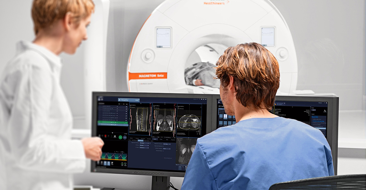
Elliot at River’s Edge utilizes a 3Tesla high field MR system to obtain all breast imaging and perform all breast MR Guided Biopsies.

After you have changed and your IV has been started, someone from the interventional team will sit down to go over the post procedure instructions. The Radiologist performing your exam will also explain the procedure including benefits and risks and have you sign consent.

The MRI Guided Biopsy requires a miniature version of the diagnostic MRI you had recently. Again this exam requires contrast and your IV will be used to administer a contrast agent called Gadavist.

Once in the scan room you will be asked to lie on your stomach onto our dedicated breast coil which was used for your diagnostic exam. The breast of concern will be placed in compression and a grid device will be placed by the Radiologist to help aid with locating the area of interest.

The procedure will take approximately 1 Hour from start to finish. We will be taking a series of pictures to determine the area of interest and then to confirm the location of the biopsy device. All of the series will be a combination of individual scans lasting only 1 minute.

The images obtain during the MRI will be sent to a special CAD system located in the control room where a Radiologist dedicated to Breast MRI will determine coordinates to locate the area of interest.

A breast tissue marker made of titanium (nonferrous material) will be placed in the breast in the area where the biopsy was performed. After the procedure is complete the interventional team will assist holding pressure on the breast and apply a dressing.

After the procedure is over you will be taken to the Mammography department for a gentle mammogram to document the clip placement.

NOTE: You are welcome to bring loved ones with you for support. They can wait in our waiting room until the test is complete.

If you have been booked for an MRI with Perfusion you will be asked to arrive at the MRI 30 minutes prior to your appointment time. The Perfusion technique is done by obtaining a Brain MRI with and without contrast. The contrast agent used by Elliot MRI is called Gadavist and requires that an IV be started in either your arm or hand.

MRI Safe clothing will be provided for you. You will be asked to lie on your back onto an MRI coil (camera) and an additional coil will be clicked in place acting as an antenna. This device has prism technology which allows you to see outside of the machine, either looking at the Technologists or the picture window depending on the site you are scanned at. You will be given hearing protection as the MRI machine makes a series of loud noises. You will also have the option of listening to satellite radio.

During the contrast portion of the MRI timed pictures will be acquired to enable the Radiologist to see color mapped images as well as time point curves. These specialized scans will be used in combination with your other brain images and a formal report of the results will be sent to your referring physician. Your MRI results will be explained to you by your referring physician.

If you have been booked for an MRCP you will be asked to arrive at the MRI department 15 minutes prior to your appointment time. It is necessary to abstain from having anything to eat or drink for 6 Hours prior to your MRCP. The MRCP technique is done by using the MRI machine and a coil (camera) to obtain images of your abdomen and biliary structures. The majority of MRCP exams DO NOT require a contrast agent. Occasionally if the MRCP is to be done in additional to an MRI Abdomen with and without contrast we will be using a contrast agent called Gadavist.

MRI Safe clothing will be provided for you to wear. You will be asked to lie on your back onto an MRI coil (camera) and an additional coil will be placed over your abdomen acting as an antenna. You will be given ear plugs or headphones (Both if you are scanned at River’s Edge location) to protect your hearing as the MRI machine makes a very loud noise.
During the scanning portion of the MRI you will be given instructions to hold your breath. The Technologist will be in constant contact to help guide you through the exam and instruct you of when to hold your breath and when to breathe. After your MRCP is done you are able to resume normal daily activities without restrictions.

If you have been booked for an MRA you will be asked to arrive at the MRI department 15 minutes prior to your appointment time. The MRA technique is done by using the MRI machine and a coil (camera) to obtain images of your arteries and/or Veins. Images of the veins are referred to as an MRV (Magnetic Resonance Venogram). The majority of MRA exams use a contrast agent called Gadavist and requires that an IV be started in either your arm or hand.

MRI safe clothing will be provided for you. You will be asked to lie on your back onto an MRI coil (camera) and an additional coil will be clicked in place acting as an antenna. You will be given ear plugs or headphones to protect your hearing as the MRI machine makes a very loud noise.

Timed images will be acquired which enable the Radiologist to visualize the vessels. You will receive the results of your MRI from your referring Physician.

If you have been booked for an MRI and you have metal in the area of concern you may be a candidate for WARP software. WARP software is a type of technology the Elliot Hospital and Londonderry MRI has built into their sequences that allow for metal artifact reduction (also known as MARS).

The technology is popular for imaging of the shoulder, hips, knees, and spine but can be used for any body part that contains metal.

If you have a knee replacement, hip replacement, shoulder replacement or metal from a fusion in your spine you may be a candidate for WARP software to better visualize the area of concern. Your referring provider can help you set up this appointment through our central booking department.

The length of an MRI with WARP is anywhere between 25-35 minutes and will require you to hold very still. MRI scans are sensitive to motion and any motion may obscure information the Radiologist uses to make a formal report. The metal you have in the area of concern will still be visible however the software will drastically reduce the artifacts seen with traditional MRI scans.

The formal report of the results will be sent to your referring physician who will explain the results to you.

Pediatric MRI Preparation

If your child is receiving an MRI, please view our helpful preparation guides for a pediatric MRI along with an MRI coloring page to help ease your child's nerves.

Pediatric Sedated MRI Preparation Information

Pediatric Non-sedated MRI Preparation Information

Download the MRI Coloring Page

ACR-Accredidtation_MRI.png


All of The Elliot MRI sites are accredited by the American College of Radiology

The Elliot encourages you to explore the below videos for more patient and safety information regarding MRI exams and our Siemens technology.

Understanding MRI (English)

Understanding MRI (Spanish)